Fluorescence microscopy is one of the most fascinating and powerful techniques in modern science. Unlike traditional light microscopes that rely on simple illumination, fluorescence microscopes reveal structures and details that would otherwise remain invisible. By using special fluorescent dyes or naturally fluorescent molecules, this method produces images with bright, glowing colors against dark backgrounds creating both stunning visuals and valuable scientific insights.
Today, fluorescence microscopy is essential in biology, biotechnology, material science, and education. It opens a new window into the microscopic world, making it a favorite among science enthusiasts, students, and researchers.
What Makes Fluorescence Microscopy Special?
Fluorescence microscopy is different from regular observation because it highlights specific structures based on how they emit light. Read more
Instead of simply shining light on a sample, it uses excitation light to make certain molecules emit fluorescent colors.
This unique process allows users to see:
Patterns
Shapes
Structures
Interactions
that are invisible under conventional light.
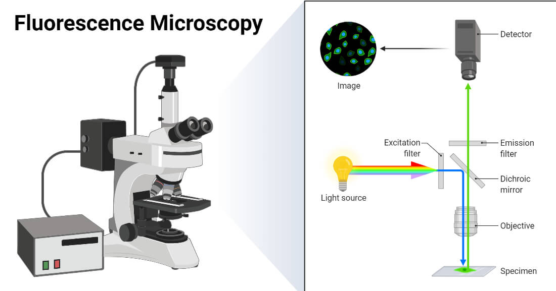
How Does Fluorescence Microscopy Work?
The principle behind fluorescence is simple yet impressive:
The microscope shines a specific wavelength of light on the sample.
Certain molecules absorb this light.
They emit light of a different color (usually brighter) that the microscope captures.
As a result, only targeted components glow, while the rest remains dark.
This contrast creates incredibly clear and precise images.
Why Is Fluorescence Microscopy So Popular?
Fluorescence microscopy has become extremely popular in recent years because it offers benefits that no other light-based microscopy technique can match.
High contrast and clear visualization
Structures appear bright against a dark background, making details easier to observe.
Specific targeting
Fluorescent dyes can bind to particular components, allowing users to highlight exactly what they want to study.
Perfect for modern education
Teachers and students love the colorful, glowing images that make microscopic structures easier to understand.
Widely used in biotechnology and research
From cell components to materials, fluorescence reveals features that other microscopes cannot capture.
Where Is Fluorescence Microscopy Used?
Fluorescence microscopy is versatile and widely applied in many fields.
Biology and Life Sciences
Used to observe:
Plant tissues
Microorganisms
Cell shapes and patterns
Natural fluorescent pigments
Biotechnology and Research
Perfect for advanced studies requiring precise visualization of structures and interactions.
Material Science
Helps highlight specific components in plastics, fibers, coatings, and pigments.
Education and Training
Popular in classrooms and teaching labs, making lessons more interactive and visually engaging.
The Visual Appeal of Fluorescence Microscopy
One of the reasons this technique is so admired is the beauty of the images it produces. Read more
Fluorescent samples often glow in:
Bright greens
Blues
Reds
Yellows
These vibrant images are extremely popular on social media and science blogs. Students and hobbyists enjoy sharing:
Glowing plant tissues
Fluorescent crystals
Illuminated microorganisms
The aesthetic aspect of fluorescence microscopy inspires curiosity and creativity.

Why Fluorescence Microscopy Feels “Like Seeing the Invisible”
Because this technique highlights specific components with glowing colors, it lets the observer see details that are:
too small
too transparent
or too faint
to be detected by regular light.
It transforms invisible structures into bright and recognizable shapes—revealing a world normally hidden from view.
Conclusion
Fluorescence microscopy is more than just a technique it’s a captivating way to explore the hidden beauty of microscopic structures. Its combination of scientific precision and visual appeal makes it one of the most exciting tools in modern microscopy.
For students, hobbyists, and researchers, it unlocks a whole new dimension of observation truly allowing us to see the invisible.
Discover our new best-seller.
Confocal Gentaur Unit